Full Name
Rudolf Oldenbourg
Title
Senior Scientist

- Email:
- Phone:
- Fax:
- CV:
- ORCID ID:
0000-0003-1055-8692
Diplomphysiker (Master in Physics), Technical University Munich, Germany, 1976
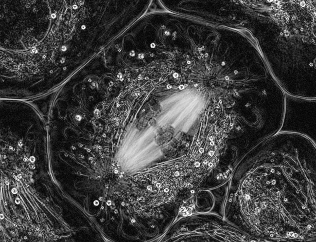
Our research is inspired by advanced optical methods for studying the architectural dynamics in living cells. We are developing polarized light microscopy techniques, including the LC-PolScope, for the analysis of dynamic molecular order directly in living cells with unprecedented sensitivity, resolution, and speed. Based on polarization measurements, we gain insight into submicroscopic structural parameters and non-invasively create contrast where native structures are otherwise invisible. We seek interdisciplinary collaborations to conduct research in physical optics for the interpretation of image content and in computational methods for image enhancement and restoration. These physical and engineering projects are stimulated and guided by biological inquiries into the structural basis of cell function.
LAB PROJECTS:
- OpenPolScope
- Polarized Light Field Microscopy
- Imaging Innovation Initiative


Koike-Tani, M., Tominaga, T., Oldenbourg, R. and Tani, T. (2020). Birefringence Changes of Dendrites in Mouse Hippocampal Slices Revealed with Polarizing Microscopy. Biophys. J., 118(10):2366-2384. http://dx.doi.org/10.1016/j.bpj.2020.03.016
Colon-Ramos, D. A., La Riviere, P., Shroff, H. and Oldenbourg, R. (2019). Transforming the development and dissemination of cutting-edge microscopy and computation. Nat Methods, 16(8):667-669. http://dx.doi.org/10.1038/s41592-019-0475-y
Chandler, T., Mehta, S., Shroff, H., Oldenbourg, R., and La Riviere, P. J. (2017). Single-fluorophore orientation determination with multiview polarized illumination: modeling and microscope design. Opt Express, 25(25):31309-31325. http://dx.doi.org/10.1364/OE.25.031309
McQuilken, M., Jentzsch, M. S., Verma, A., Mehta, S. B., Oldenbourg, R., and Gladfelter, A. S. (2017). Analysis of Septin Reorganization at Cytokinesis Using Polarized Fluorescence Microscopy. Front Cell Dev Biol, 5:42. http://dx.doi.org/10.3389/fcell.2017.00042
Tani, T., M. Shribak and R. Oldenbourg. 2016. Living Cells and Dynamic Molecules Observed with the Polarized Light Microscope: the Legacy of Shinya Inoue. Biological Bulletin 231: 85-95. http://dx.doi.org/10.1086/689593
Abrahamsson, S., M. McQuilken, S. B. Mehta, A. Verma, J. Larsch, R. Ilic, R. Heintzmann, C. I. Bargmann, A. S. Gladfelter and R. Oldenbourg. 2015. MultiFocus Polarization Microscope (MF-PolScope) for 3D polarization imaging of up to 25 focal planes simultaneously. Optics Express 23: 7734-7754. https://doi.org/10.1364/OE.23.007734
Oldenbourg, R. 2008. Polarized light field microscopy: an analytical method using a microlens array to simultaneously capture both conoscopic and orthoscopic views of birefringent objects. J. Microsc. 231: 419-32. https://doi.org/10.1111/j.1365-2818.2008.02053.x
Oldenbourg, R., E. D. Salmon and P. T. Tran. 1998. Birefringence of single and bundled microtubules. Biophys. J.74: 645-54. https://doi.org/10.1016/S0006-3495(98)77824-5
Oldenbourg, R. 1996. A new view on polarization microscopy. Nature 381: 811-2. https://doi.org/10.1038/381811a0
Oldenbourg, R. and G. Mei. 1995. New polarized light microscope with precision universal compensator. J. Microsc.180: 140-7. https://doi.org/10.1111/j.1365-2818.1995.tb03669.x
Oldenbourg, R., X. Wen, R. B. Meyer and D. L. Caspar. 1988. Orientational distribution function in nematic tobacco-mosaic-virus liquid crystals measured by x-ray diffraction. Phys. Rev. Lett. 61: 1851-1854. https://doi.org/10.1103/PhysRevLett.61.1851