Cell Specialization
As the 19th century progressed, new methods emerged to fix, slice, and stain tissues and therefore interpret their constituent cells. Especially within medicine, it became clear that cells organize into specialized tissues and organs with varying shapes, playing different roles within a larger organism.
Gradually, with the emerging field of cytology, researchers began to focus on one type of cell or another, exploring in greater detail their structure and function.
 HoverTouch to magnify
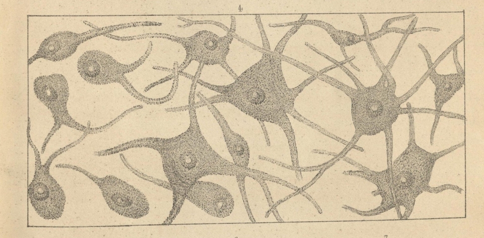
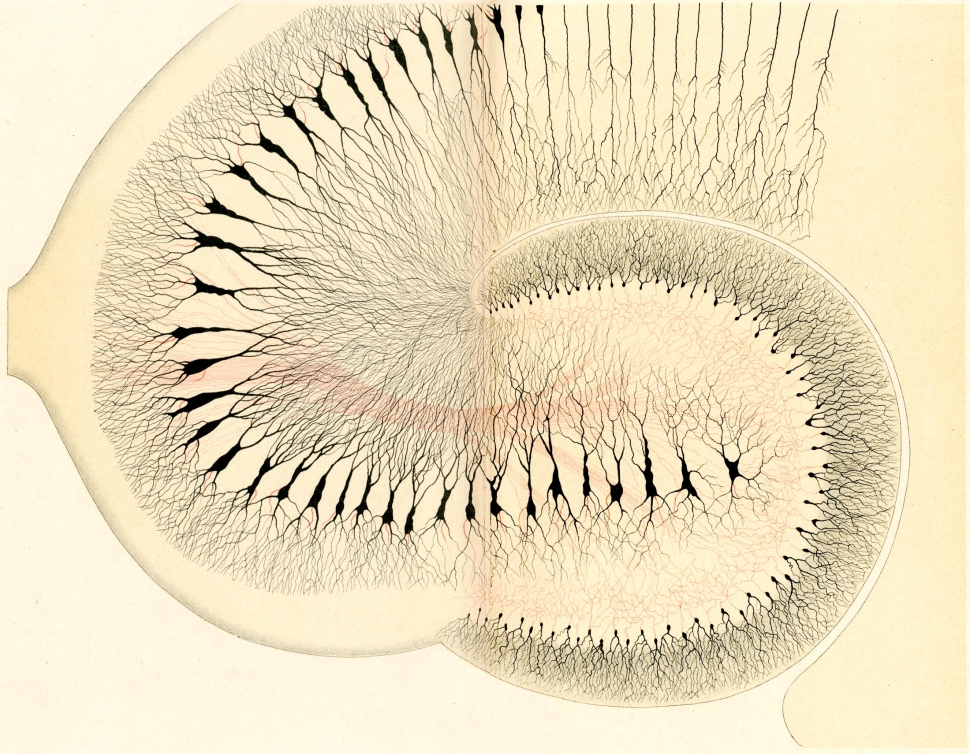
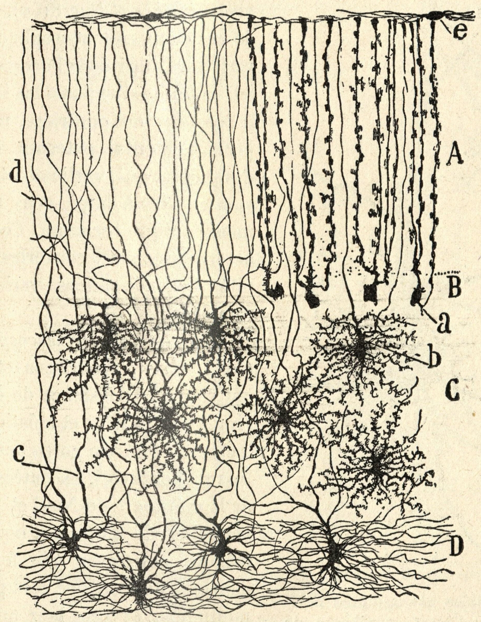
For example, with nerve cells: Italian microscopist Camillo Golgi and Spaniard Santiago Ramón y Cajal used new staining methods to explore nerve cells and how they group and work together in nervous systems. Although they looked at much the same things, they vehemently disagreed in their interpretations. Where Ramón y Cajal saw separate nerve cells, Golgi saw a single connected mass called a reticulum.
 HoverTouch to magnify
 HoverTouch to magnify
They shared a Nobel Prize but still disagreed. Each pointed to his particular images as evidence for his claims. Their innovations, observations, images, and arguments show how exciting this time in history was. And how difficult it can be to interpret what we see with microscopes.
- Hassall, Arthur Hill. The Microscopic Anatomy of the Human Body in Health and Disease. London: Taylor, Walton, and Maberly, 1852. Plate XLV.
- Golgi, Camillo. “Sulla fina Anatomia Degli Organi Centrali Del Sistema Nervoso, Rivista sperimentale di Freniatria, anno 1883.” 399-539. Opera Omina, Volume II Istologia Normale 1883-190, Ulrico Hoepli, Editore Libraio Della Real Casa Milano 1903. Page 532, Table XXXI.
- Ramón y Cajal, S. Histologie du Système Nerveux de l'Homme & des Vertébrés. Paris: A. Maloine, 1909. Page 237, Figure 81.