Seeing How Cell Parts Move
By the 1970s, electron microscopy and live cell imaging showed that cells are filled with different types of very fine filaments, like microtubules, that form a cytoskeleton.
Microtubules were too small to see with ordinary light microscopy. But while developing a video microscope at the MBL, Nina and Robert Allen accidentally discovered that they could detect cellular structures below the resolution of light microscopy by artificially increasing the contrast of their live cell images. Shinya Inoué, also at the MBL, almost simultaneously designed a similar video microscope.
Together with Scott Brady and Ray Lasek, who worked summers at the MBL on a nerve cell process from squid called the giant axon, Robert Allen visualized tiny vesicles moving along microtubules within the axon.
 HoverTouch to magnify
Brady and Ron Vale were soon using the same video setup to view vesicles moving on microtubules isolated from the axon.
 HoverTouch to magnify
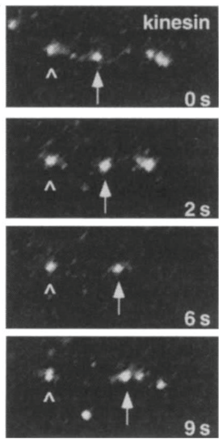
Biochemical analysis of the cell-free preparation led Vale to discover a new molecular motor which he called kinesin.
 HoverTouch to magnify
- Allen, Robert Day, Nina Stromgren Allen, and Jeffrey L. Travis. 1981. "Video-Enhanced Contrast, Differential Interference Contrast (AVEC-DIC) Microscopy: A New Method Capable of Analyzing Microtubule-Related Motility in the Reticulopodial Network of Allogromia laticollaris." Cell Motility 1: 291-302. Page 297, Figure 4.
- Vale, Ronald D., Bruce J. Schnapp, Thomas S. Reese, and Michael P. Sheetz. 1985. "Movement of Organelles Along Filaments Dissociated from the Axoplasm of the Squid Giant Axon." Cell 40: 449-454. Page 450, Figure 1.
- Vale, Ronald D, Takashi Funatsu, Daniel W Pierce, Laura Romberg, Yoshie Harada, and Toshio Yanagida. 1996. "Direct Observation of Single Kinesin Molecules Moving Along Microtubules." Nature (London) 380(6573): 451–453. Page 451, Figure 1.