Video Microscopy
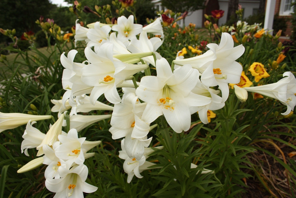
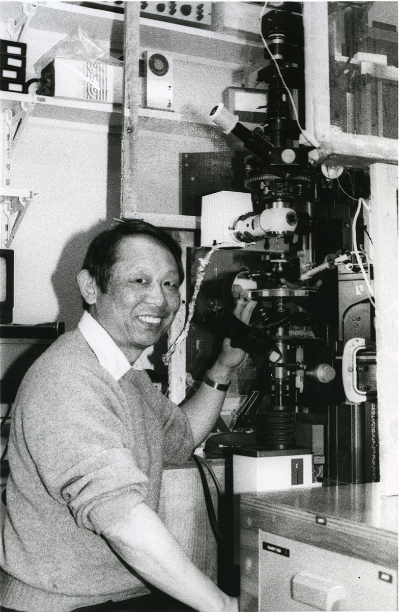
Adding to his abilities in microscope building and his unique approach to understanding mitotic spindles, Inoué also understood the power of moving images to portray his findings. In the summer of 1951, Inoué showed a time lapse movie of Lilium spindle fibers diving, which he captured with his second version of the Shinya-scope.
 HoverTouch to magnify

 HoverTouch to magnify
The collaborative atmosphere among faculty, students, and the representatives of relevant industries participating in the courses established Video Microscopy as an important research tool in cell biology. In addition, the course curricula soon included an important next step in image enhancement and quantitation made possible by the availability of desktop computers with video frame grabbers, opening up with the new and vast field of digital imaging and microscopy.
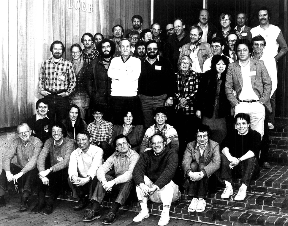
In 1980, Inoué started the Analytical & Quantitative Light Microscopy (AQLM) course at the MBL alongside Bob Allen’s Optical Microscopy course. While using video to display live images from the microscope in the classroom, they realized that adjusting the brightness and contrast of the images on the video equipment allowed them to improve the visibility of details too faint to be seen under normal viewing conditions.
1. Wikimedia Commons contributors, "File:Easter Lily Cluster in Garden.jpg," Wikimedia Commons, the free media repository, https://commons.wikimedia.org/w/index.php?title=File:Easter_Lily_Cluster_in_Garden.jpg&oldid=563463820 (accessed October 11, 2021).
2. Still Image: "Shinya Inoue with Microscope", 6/12/17 https://hdl.handle.net/1912/22636.
3. Still Image: "Analytical and Quantitative Light Microscopy Course Photo in 1983", 7/7/2015, https://hdl.handle.net/1912/21888.