 HoverTouch to magnify
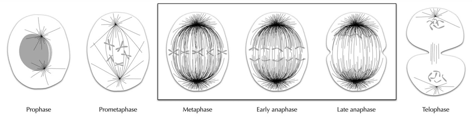
Animal cell division
These illustrations show Inoué’s studies of the mitotic spindle during cell division. He used birefringence, so he mainly saw mitotic spindles and less of other cell elements.
 HoverTouch to magnify
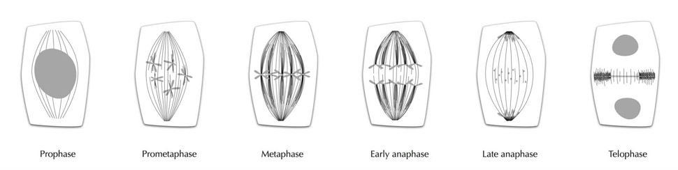
Plant cell division
These drawings depart from classical drawings of cell division in several ways: First, the nucleus, chromosomes, and cell membranes are in grey to highlight the mitotic spindles. Second, centrioles and centromeres were not visible in Inoue’s images, so they are not drawn. Third, the phases that were of particular importance for Inoué are drawn in more detail.
1. Still Image: "Stages of cell division in animals and plants by Lucie Laplane", 7/7/2015, https://hdl.handle.net/1912/21901.