Resolving the Inside of Cells
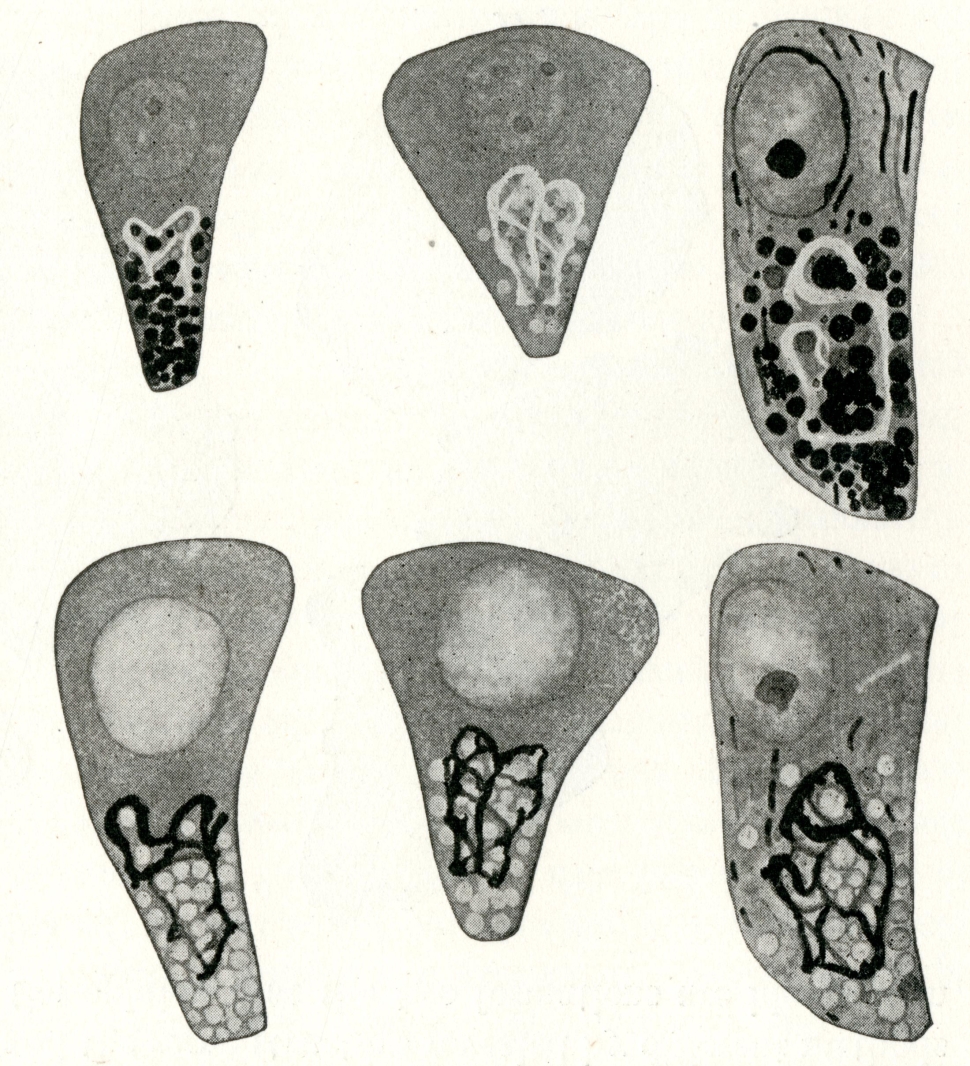
Before 1900, it was not clear which structures that appeared to exist inside cells were real and meaningful for the cell. Maybe some were artifacts of the way the cells had to be prepared for observation with the light microscope. And maybe other things existed that were not visible microscopically.
 HoverTouch to magnify
 HoverTouch to magnify
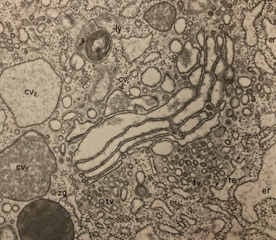
In the 1930s and 1940s, use of the electron microscope (EM) to in biology generated new ways to access cellular anatomy. EM made it possible to see more by seeing differently. The greatly increased resolution compared to light microscopy helped researchers visualize the internal parts, or organelles, in much finer detail and to discover new structures in what had been previously deemed the “optically empty” part of the cytoplasm.
 HoverTouch to magnify
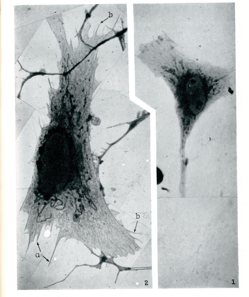
Electron micrograph resolving previously invisible structures (left). Light micrograph could not resolve the structure of the endoplasmic reticulum (right).
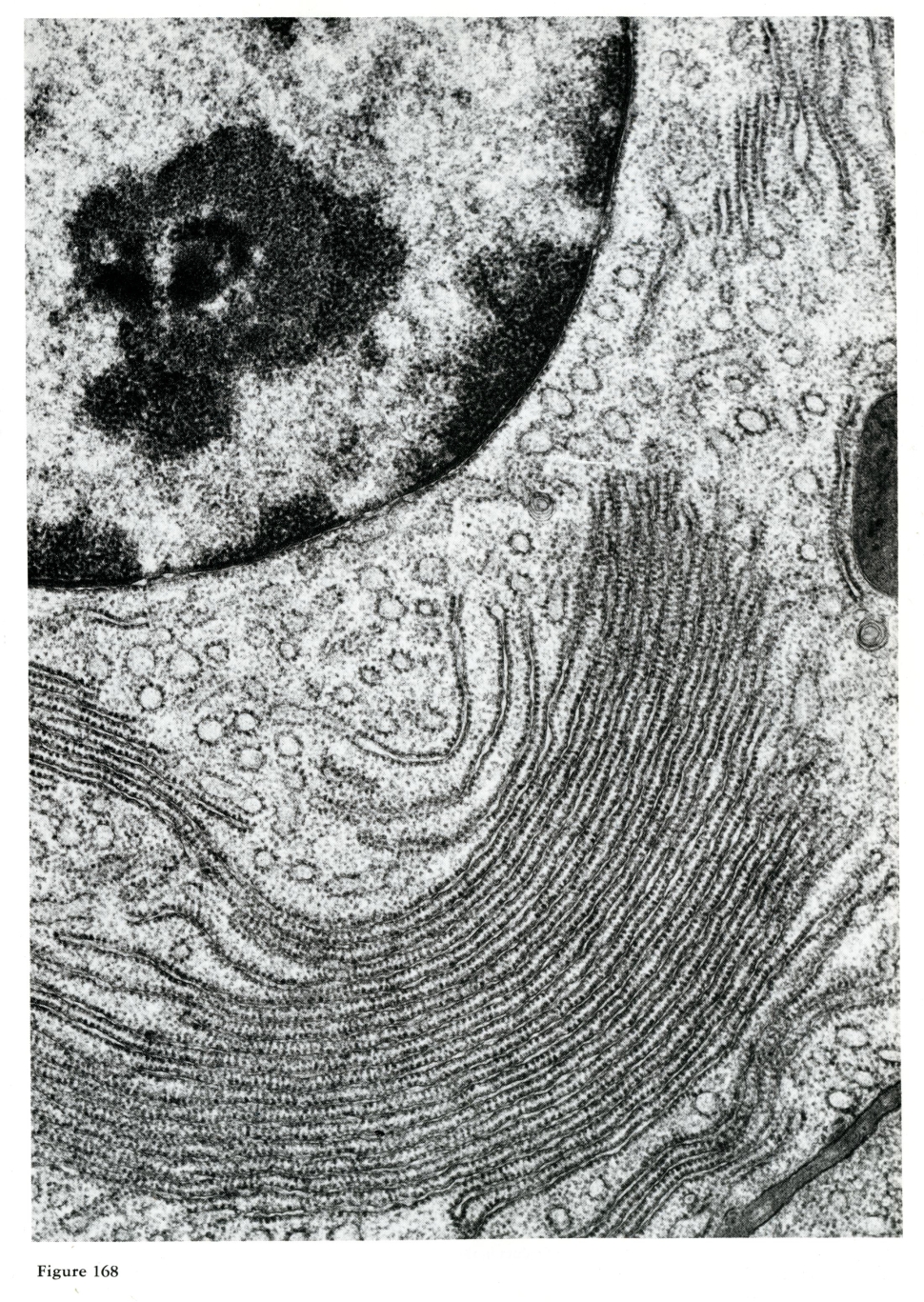
Further improvements in electron microscopy resulted in even more magnified and detailed images, which influenced theories about organelle function. EM brought new revelations and questions, while also producing exciting new images that required interpretation.
 HoverTouch to magnify
- Farquhar, Marilyn Gist, and George E. Palade. 1981. “The Golgi Apparatus (Complex) -- from Artifact to Center Stage.” The Journal of Cell Biology 91 (1): 77s-103s. Page 82s, Figure 7.
- Porter, Keith R., Albert Claude, and Ernest F. Fullam. 1945. “A Study of Tissue Culture Cells by Electron Microscopy.” Journal of Experimental Medicine 81 (3): 233-246. Page 246, Plate 10, Figures 1 and 2.
- Cowdry, Edmund V. General Cytology: A Textbook of Cellular Structure and Function for Students of Biology and Medicine. Chicago: The University of Chicago Press, 1924. Page 342, Figure 30.
- Fawcett, Don W. The Cell. Philadelphia: W. B. Saunders Co, 1981. Page 311, Figure 168.