From Fertilization to Organisms
From the MBL’s beginning in 1888, director Charles Otis Whitman inspired cell lineage studies. MBL researchers studied how a single fertilized egg becomes a multicellular embryo and on to an organized complex organism.
 HoverTouch to magnify
 HoverTouch to magnify
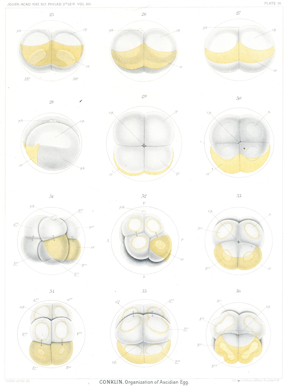
Using different marine organisms, they followed the lineage through every cell division, watching fertilized egg cells divide into 2, 4, 8 cells, and so on. Fixing, staining, observing, drawing, and photographing allowed each researcher to share and compare with others. The images began to reveal the patterns of development.
 HoverTouch to magnify
Comparing embryos from different species showed that cells can divide faster or slower, into larger or smaller cells, sometimes through spiral cleavage, and always in ways that cause differentiation among cells that allows them to form organisms.
 HoverTouch to magnify
Successive cleavage stages in slipper nail embryos from 4 cells (top) to 8 cells (bottom).
Photomicrographs with fluorescently labeled organelles (right).
(left) Conklin 18974
(right) Henry & Lyons 20145
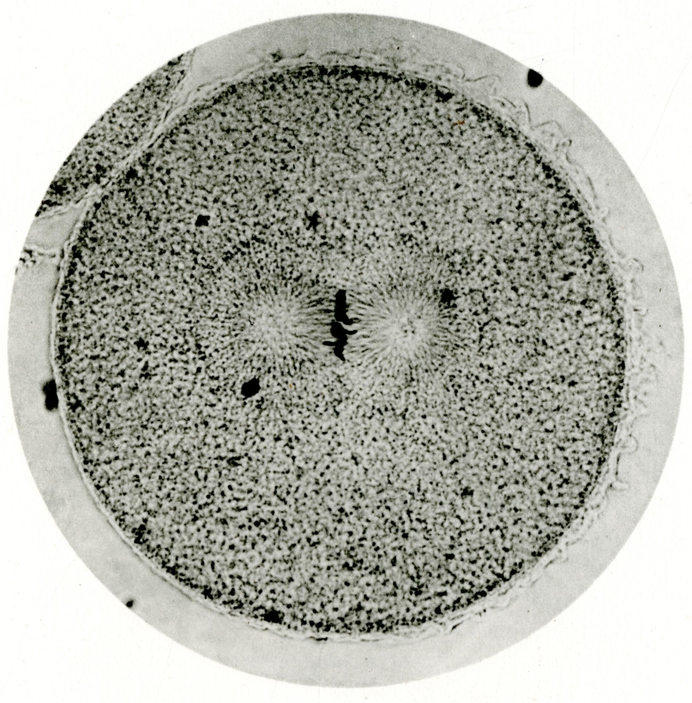
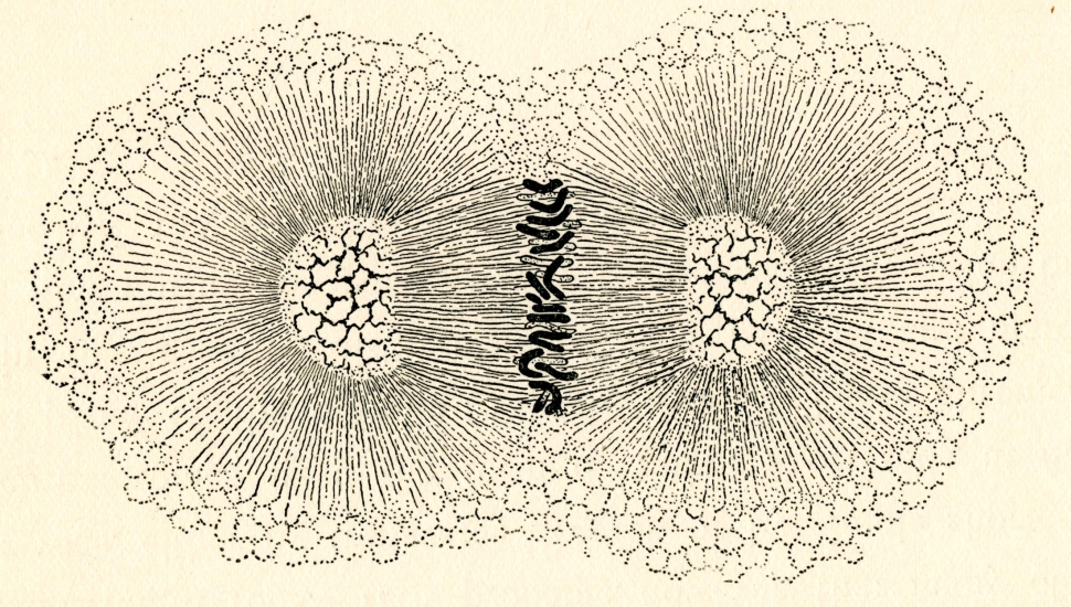
As they watched the cells divide into many hundreds and even thousands more cells and differentiate, they began to see new cell parts: a nucleus with chromosomes, spindle fibers to help direct the dance of cell division, organelles, and other details.
- Wilson, Edmund Beecher. An Atlas of the Fertilization and Karyokinesis of the Ovum. New York: Columbia University Press by Macmillan and Company, 1895. Plate VII, Figure 26.
- Wilson, Edmund Beecher. An Atlas of the Fertilization and Karyokinesis of the Ovum. New York: Columbia University Press by Macmillan and Company, 1895. Plate II, Figure XIV.
- Conklin, Edwin Grant. The Organization and Cell-Lineage of the Ascidian Egg. Philadelphia: Academy of Natural Sciences, 1905. Plate III.
- Conklin, Edwin Grant. 1897. “The Embryology of Crepidula, A Contribution to the Cell Lineage and Early Development of Some Marine Gastropods.” Journal of Morphology Vol. XIII, No. 1. Plate I, Figure 12 and Plate II, Figure 13.
- Henry, Jonathan & Deirdre Lyons. The International Journal of Developmental Biology. Vol. 58, No's 6/7/8, 2014. Cover figure.