Seeing Cells in Life
The 17th century brought microscopes, which made looking at small living material possible. Observers recorded and shared observations through letters and publications, with illustrations to show others what they saw.
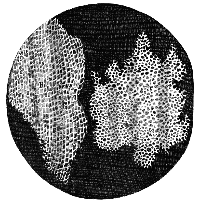
In his 1665 Micrographia, British microscopist Robert Hooke applied the term “cell” to the empty spaces he observed in slices of cork, vegetables, and sea shells. His cells were units of organization rather than living things.
 HoverTouch to magnify
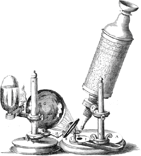
Hooke's Microscope
 HoverTouch to magnify
Cells observed in cork
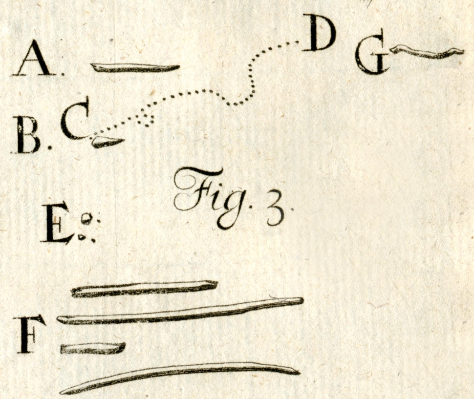
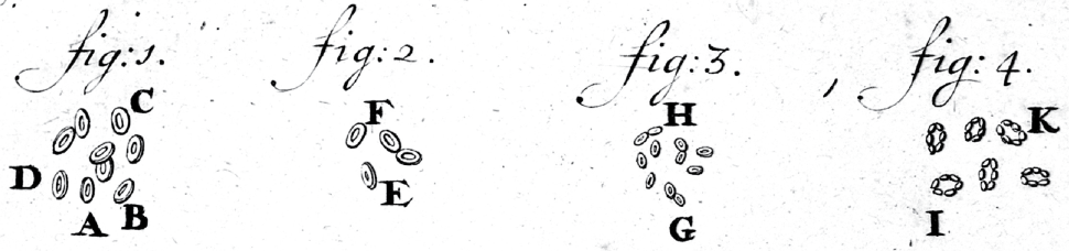
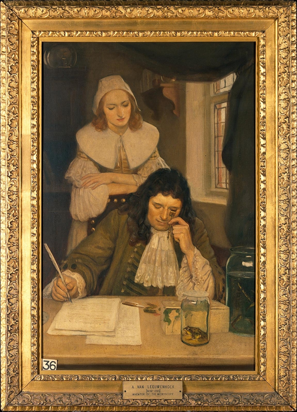
Meanwhile, in the Netherlands, Anton van Leeuwenhoek focused on living materials. He saw wiggling ovals in scrapings from his mouth and called them “animalcules.” Non-moving disks in blood he called “corpuscles.”
 HoverTouch to magnify
Dental plaque animalcules
MBL Rare Books Collection
 HoverTouch to magnify
Blood corpuscules
 HoverTouch to magnify
van Leeuwenhoek
 HoverTouch to magnify
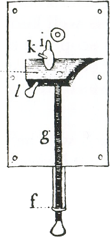
Leeuwenhoek's microscope
Others also began to see organized units of life that they called cells. They focused on observing and describing what they saw and did not yet develop a theory of cells. That came in the 19th century with improved microscopes. What was that cell theory?
- Hooke, Robert. Micrographia: or, Some Physiological Descriptions of Minute Bodies Made By Magnifying Glasses. With Observations and Inquiries Thereupon. London: Printed by J. Martyn and J. Allestry, 1665. Schem. I, Figures 5 and 6.
- Hooke, Robert. Micrographia: or, Some Physiological Descriptions of Minute Bodies Made By Magnifying Glasses. With Observations and Inquiries Thereupon. London: Printed by J. Martyn and J. Allestry, 1665. Schem. XI, Figure 1.
- Leeuwenhoek, Anton van. 1684. "An abstract of a letter from Mr. Anthony Leevvenhoeck at Delft, dated Sep. 17. 1683. Containing some microscopical observations, about animals in the scurf of the teeth, the substance call'd worms in the nose, the cuticula consisting of scales" Philosophical Transactions (14) 159: 568-574. Figure 3, A-G.
- Leeuwenhoek, Anton van. 1701. "III. Part of a letter from Mr. Lewenhoek, concerning the circulation and globules of the blood in butts." Philosophical Transactions (22) 263: 552-560. Figures 1-4.
- Baker, Henry. Employment for the Microscope. London: Printed for R. and J. Dodsley, 1753. Page 440, Plate XVII, Figure VIII.
- Board, Ernest, Leeuwenhoek With His Microscope, Oil on canvas, c. 1912, (Wellcome Collection).