Seeing Cell Aggregates
In nature, most cells are parts of interacting living systems or organisms. One way to study cells interacting is by looking at aggregates.
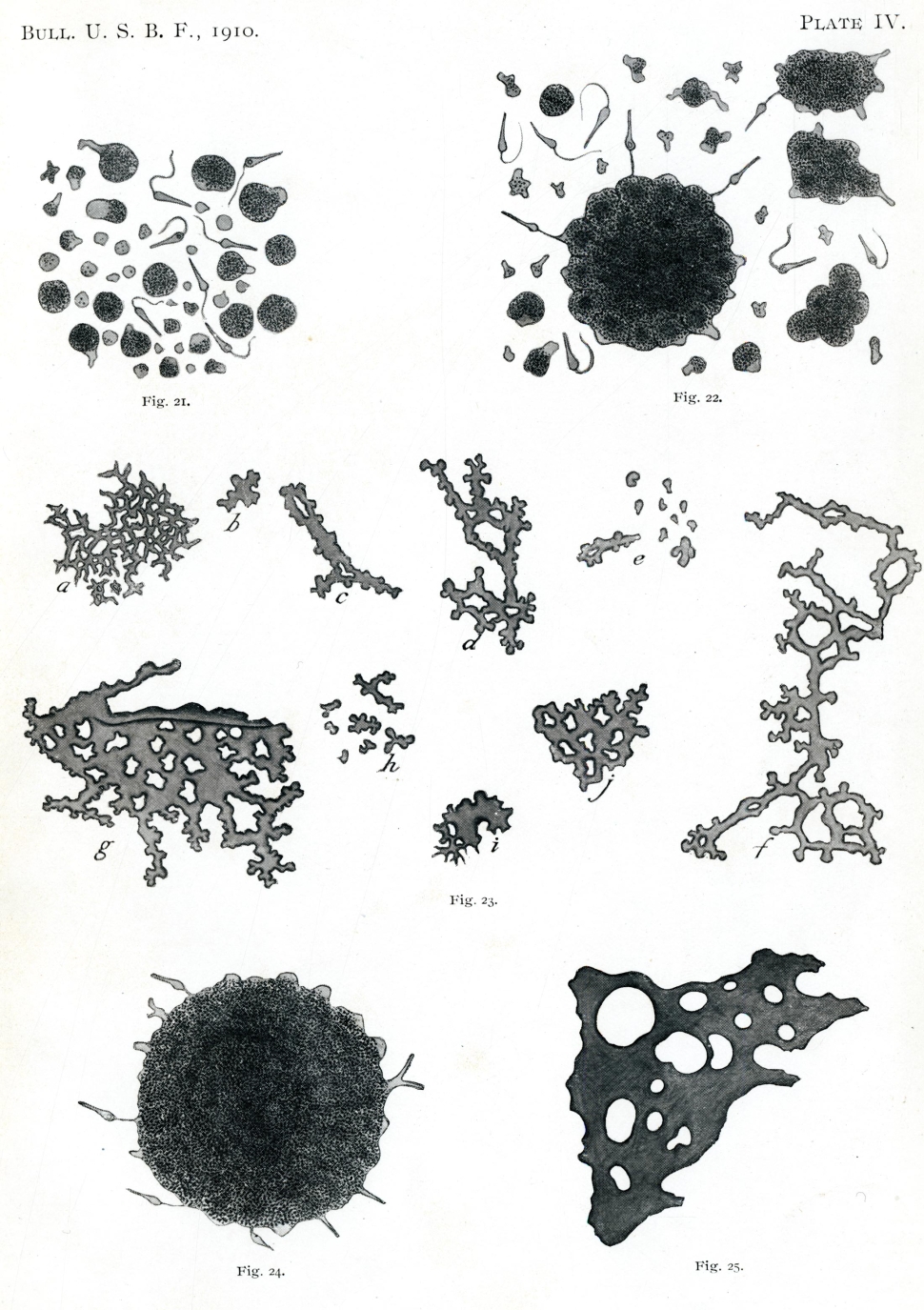
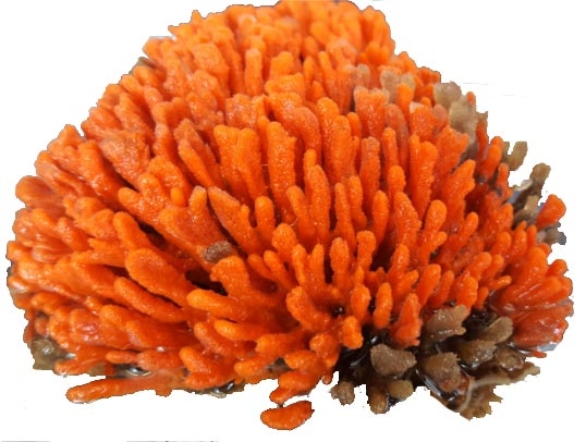
The sponge, while a multicellular organism, is considered an aggregate of individual cells. Around 1900, American Henry van Peters Wilson showed that individual sponge cells can separate and reaggregate to make living sponges again. Generations of MBL researchers have asked how this occurs.
 HoverTouch to magnify
 HoverTouch to magnify
In 1988, summer MBL researcher Gerald Weissmann asked what factors stimulate sponge cell aggregation. He put dissociated sponge cells in clear containers and found that more light led to more aggregated sponge cells. Sponge cells also need calcium to group, and other chemicals made them group even more. His work extended to show how calcium and other chemicals work in human cells. This study of aggregations gave insights into the ways that cells interact within a whole.
 HoverTouch to magnify
Seeing groups of cells at the microscopic level is not so straightforward. Researchers invent ways to tell different kinds of cells apart, and ways to interpret interactions. This has caught the attention of MBL researchers on microbial communities in particular.
 HoverTouch to magnify
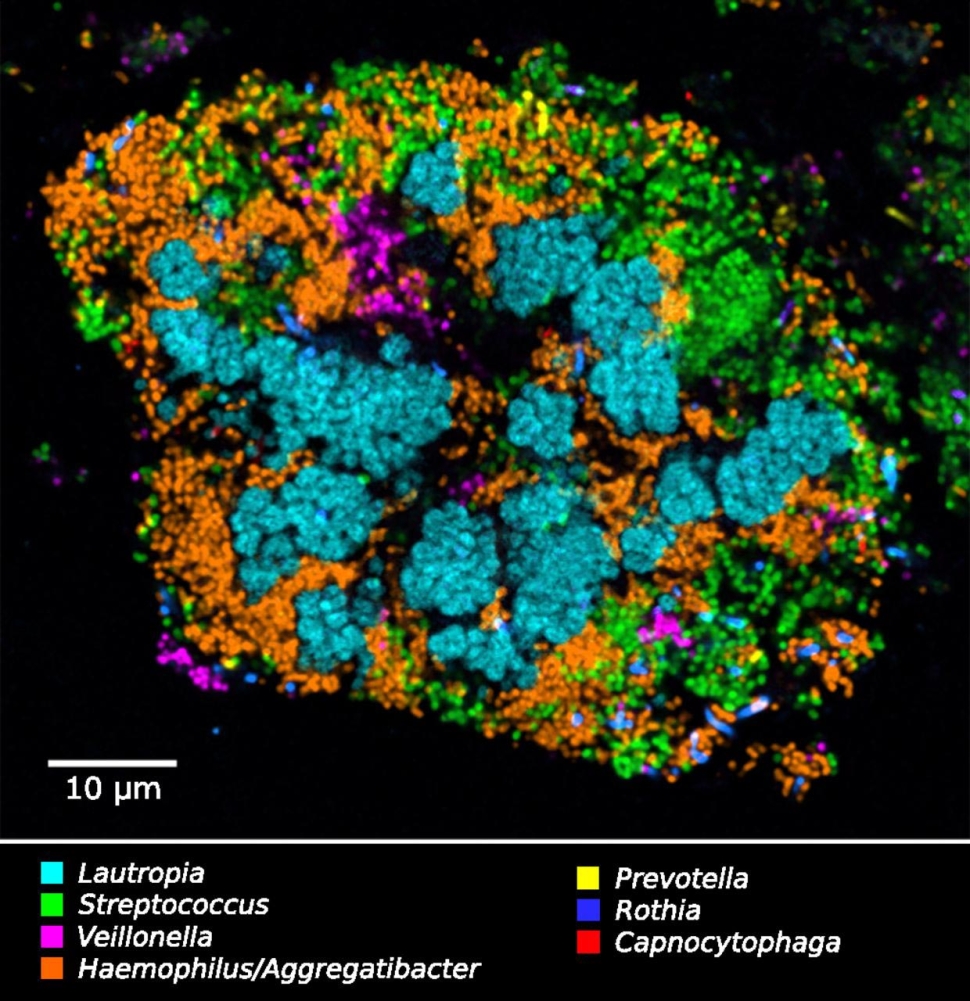
A "cauliflower" aggregate with different bacterial species labeled
One strategy to see groups of microorganisms is by making different species of microbes fluoresce specific colors. With the CLASI-FISH technique, MBL researcher Jessica Mark Welch visualizes aggregates of cells in dental plaque and learns about their spatial relationships.
- Wilson, Henry Van Peters. 1910. "Development of Sponges from Dissociated Tissue Cells." Bulletin of the Bureau of Fisheries, Volume XXX. Document N. 750, Issued June 16, 1911. Plate IV, Figure 22.
- McCallum, Monica E. July 8 to August 22, 2018. “Imaging the Microbial Community of the Marine Sponge Clathria prolifera.” Microbial Diversity Course at the Marine Biological Laboratory. Figure 1a.
- Weissmann, Gerald, Leslie B. Vosshall, Cynthia A. Bayer, and Phillip B. Dunham. 1985. “Marine Sponge Aggregation: A Model for Effects of NSAIDs on the Calcium Movements of Cell Activation.” Seminars in Arthritis and Rheumatism 15 (2): 42-53. Page 44, Figure 1A.
- Mark Welch, Jessica L., Blair J. Rossetti, Christopher W. Rieken, Floyd E. Dewhirst, and Gary G. Borisy (2016). “Biogeography of a Human Oral Microbiome at the Micron Scale.” Proceedings of the National Academy of Sciences of the United States of America 113 (6), E791-E800. Figure 8.