Inside Living Cells
In the 20th century, invention of new purely optical methods, such as differential interference contrast and phase contrast, enabled researchers to study living cells and their organelles in motion.
These inventions made observing living cells much easier. Researchers realized that cells not only move as a whole, but that organelles inside cells are constantly moving in the cytoplasm.
 HoverTouch to magnify
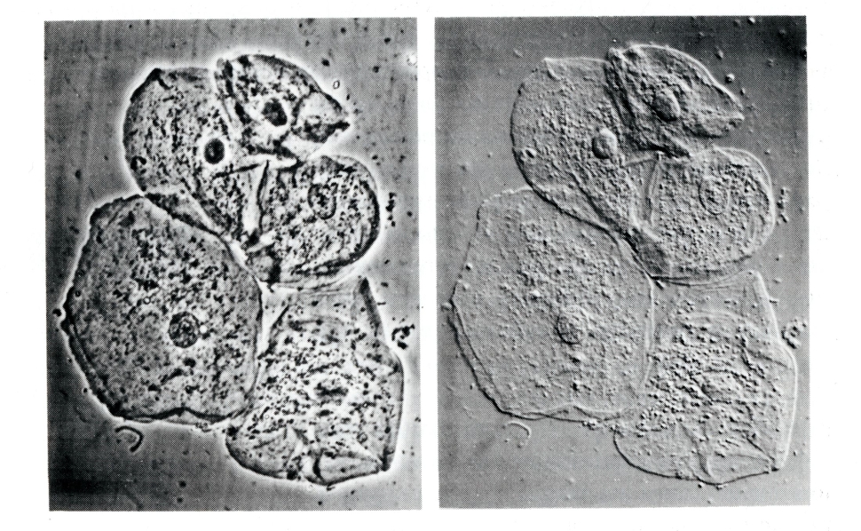
Differential interference contrast (DIC) image of oral mucus (left). Phase contrast image of oral mucus (right).
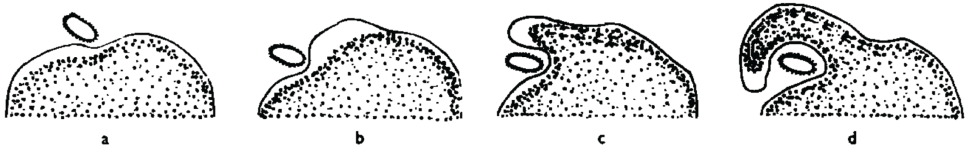
To study the mechanisms of this motion, biologists like the MBL’s Robert Allen hooked up movie cameras to his microscopes and recorded the movement of cells, like amoebae, over long time periods.
 HoverTouch to magnify
Seeing and documenting living cells in motion transformed how people thought of cells. Once static, fixed structures, cells and their parts were now visible as dynamic entities that allow changes in the cell as a whole, like cell division and differentiation.