New State-of-the-Art Microscopes Installed at MBL
Two microscopes funded by a $4.3 million award from the Massachusetts Life Sciences Center (MLSC) have been installed at the Marine Biological Laboratory (MBL). The two advanced instruments—a plasma focused ion beam-scanning electron microscope (Thermo Scientific PFIB-SEM Helios Hydra) and a confocal and super-resolution microscope (Leica Stellaris 8 STED)—will allow 2D and 3D reconstruction from both electron and light microscopy for comparative and high-resolution imaging.
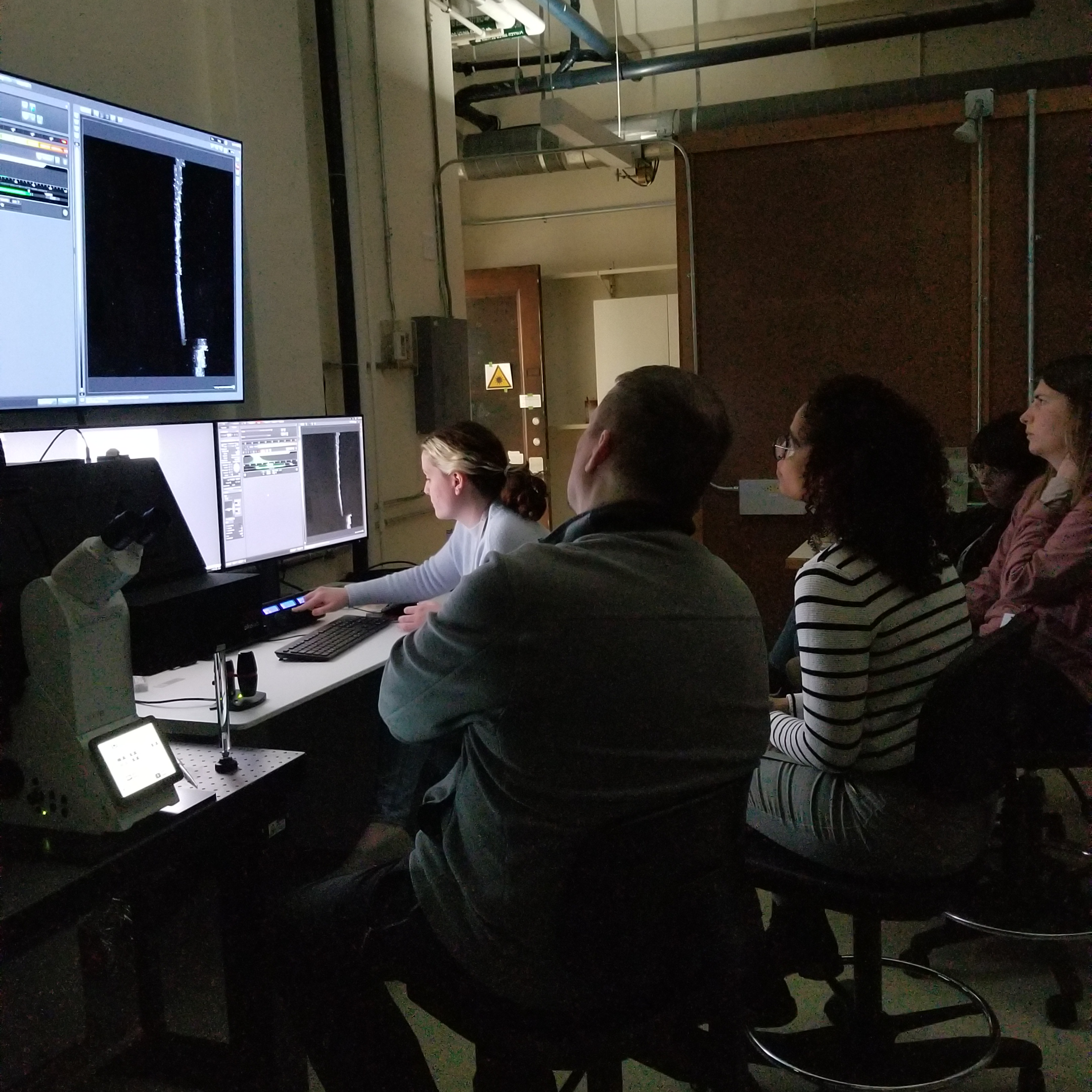
Installation began in late March and the staff of the MBL’s Central Microscopy Facility are currently fine tuning the instruments and training select staff. The facilities will be open to MBL investigators and courses along with visiting researchers from Massachusetts and beyond.
"The MBL has long been at the frontier of new imaging technologies for biological discovery. These new instruments supported by the MLSC further advance our capabilities and bring new high resolution imaging to the MBL and the broader research community,” said Anne Sylvester, MBL director of research.
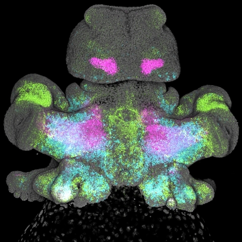
The biomedical sciences often rely on advances in imaging technologies to translate basic research into practical medical applications. Traditional 2D imaging limits the capability to study a number of physiological conditions like neurodegenerative diseases, cancer, regeneration, and wound healing. To expand into new imaging capabilities, the MBL has established an Imaging Innovation Initiative for research and training in next-generation correlative microscopy and computational image analysis that permits 3D reconstruction of biological samples. Concurrently, MBL has invested in imaging engineers and specialists who can design, build, and innovate instruments for specific use by biologists
"The MBL is excited about adding new technology to our imaging capabilities to the MBL and region," said Louis Kerr, director imaging services at the MBL. "These new instruments, which will be available to the MBL community of scientists as well as colleagues at other Massachusetts institutions, will enable imaging of biological material using light and electron microscopy and will provide new views and better resolution of biological and material samples."
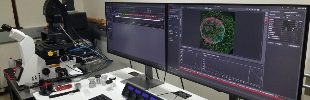
The Leica Stellaris 8 STED is a versatile high-end confocal microscope that generally provides the ability to collect clear images from a thin (optical) section of a thick sample with low background and minimal out-of-focus interference. Serial optical sectioning in combination with 3D reconstruction is a common application in the biomedical sciences. Furthermore, the Stellaris 8 includes Fluorescence Lifetime Imaging Microscopy (FLIM) that can separate different fluorophores based on their physiology and functional environment, and Stimulated Emission Depletion Microscopy (STED) which is a super-resolution technique that overcomes the diffraction limit of light by using two laser beams.
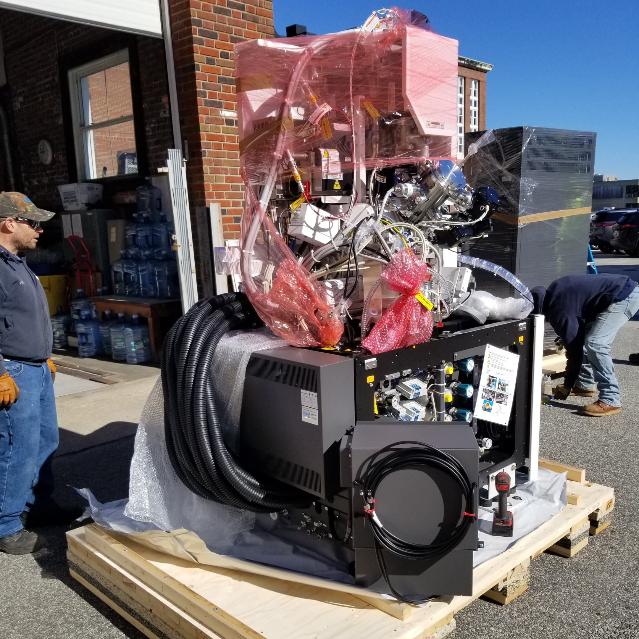
The PFIB-SEM Helios 5 Hydra is a powerful instrument that combines two key technologies: a plasma focused ion beam (PFIB) and a scanning electron microscope (SEM). The PFIB component produces a focused beam of ions (either argon, nitrogen, oxygen, or xenon), which can be precisely directed onto a sample surface. The ion beam can be used for material removal (milling) or deposition at the nanoscale. This hybrid instrument is used for standard SEM imaging, high-resolution imaging, three-dimensional (3D) reconstruction, and nanoscale material removal or modification on various sample materials at room or cryo temperatures.
Currently, there are approximately fifteen Helios Hydra systems in the U.S. and only one in Massachusetts, which is not available for public use. The microscope housed at the MBL will be the first of its kind used for biological sciences in New England. As new additions to the MBL’s Central Microscopy Facility, these advanced light and electron microscopes will provide a valued imaging resource for researchers and industry partners across the state.
Interested in utilizing either of these two new machines at the MBL? Sign up for updates and start planning for use in Summer/Fall 2024!