MBL Imagers Take Honors in Microscopy Contests

An image from the MBL's Central Microscopy Facility won first place in a prestigious microscopy contest this year, while Michael Shribak, senior scientist in the MBL's Bell Center, received recognition in three imaging contents over the past few months!
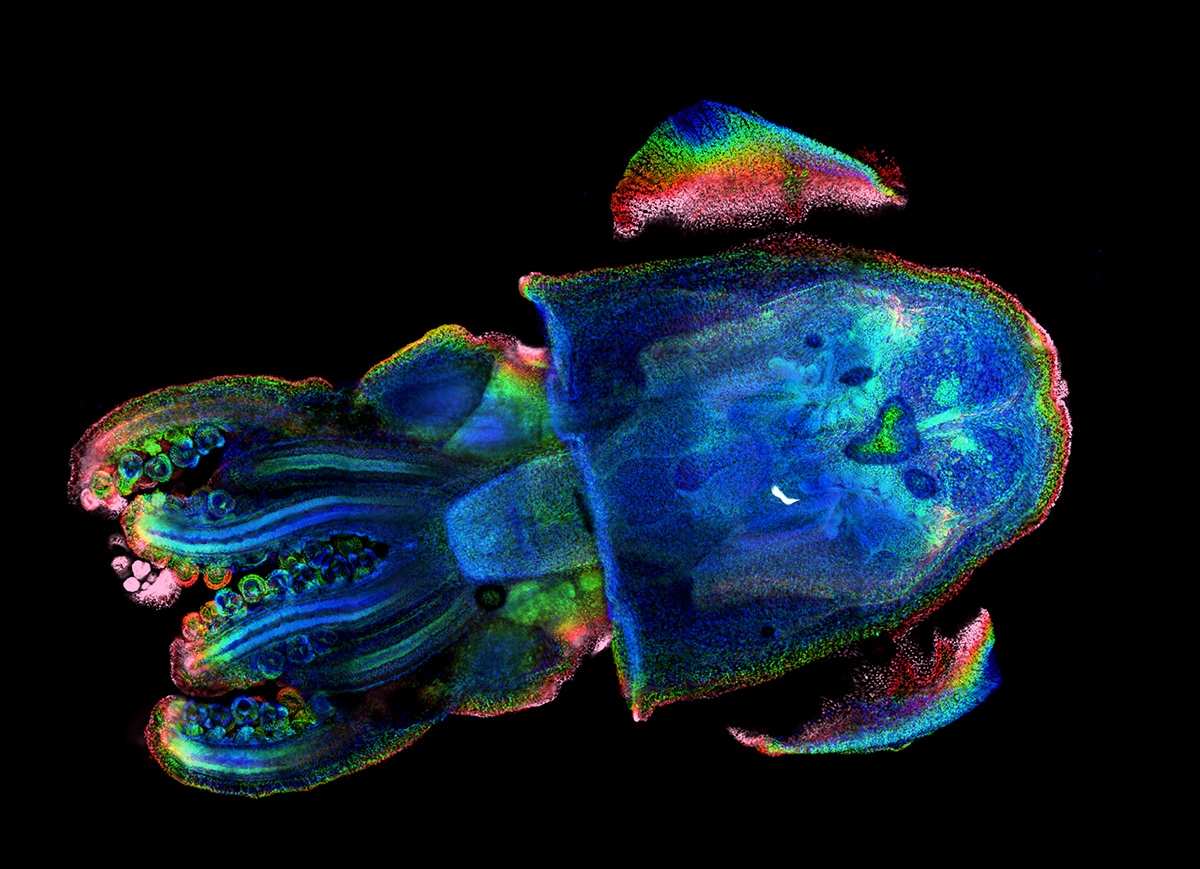
"It Takes Two to Tanglow" placed first in the BioImaging North America 2023 contest. Submitted from the MBL by Derrick Kamp of University of Connecticut, it shows the squid Euprymna berryi and its glowing symbiont, a marine bacterium:
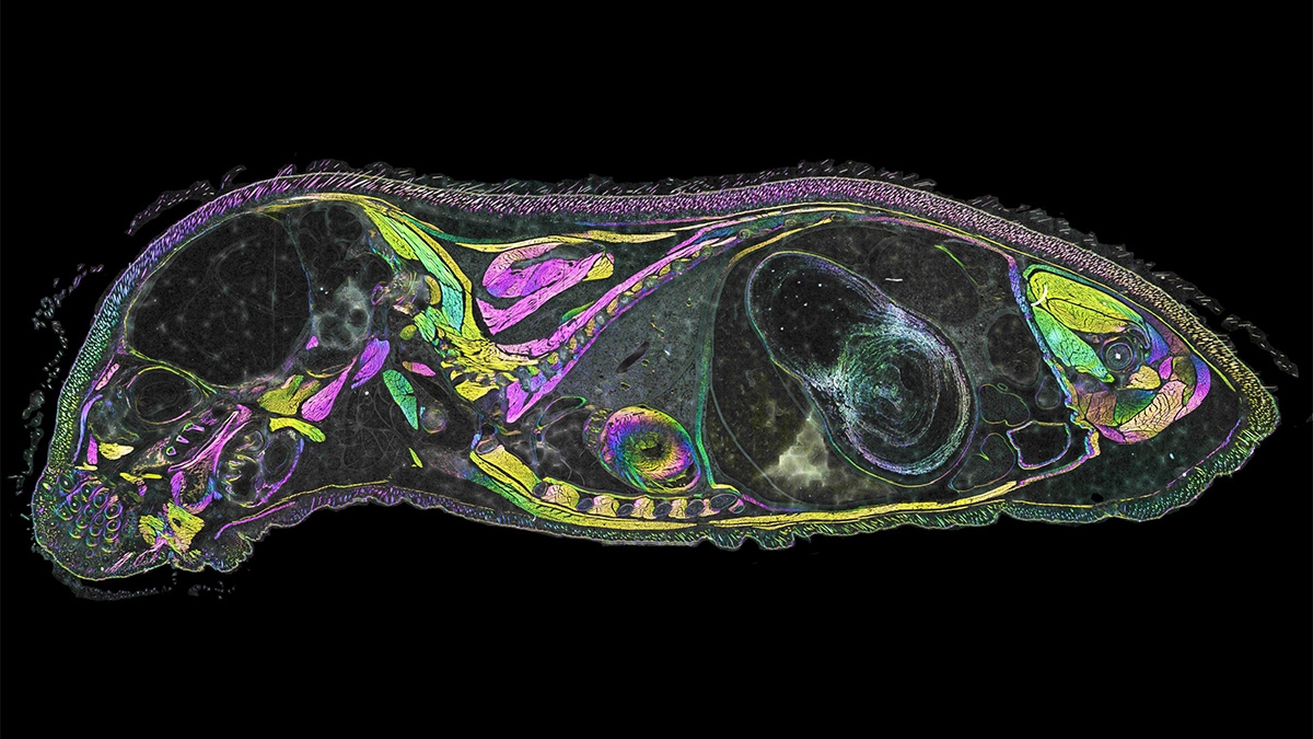
Shribak took third place in the BioImaging North America Contest for his label-free image of a mouse sagittal section (below, details and high-resolution download here). He also submitted other beautiful images, including of the diatom Actinoptychus and of stellate trichomes of oak.
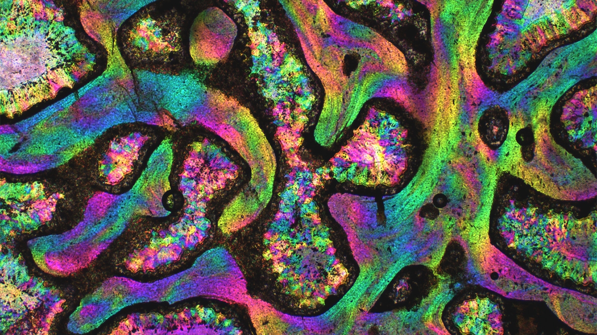
This week, Shribak learned he had placed second in the HOOKed on Microscopy Contest, sponsored by Clemson University in South Carolina, for his image, "Dinosaur Bones" (below and top).
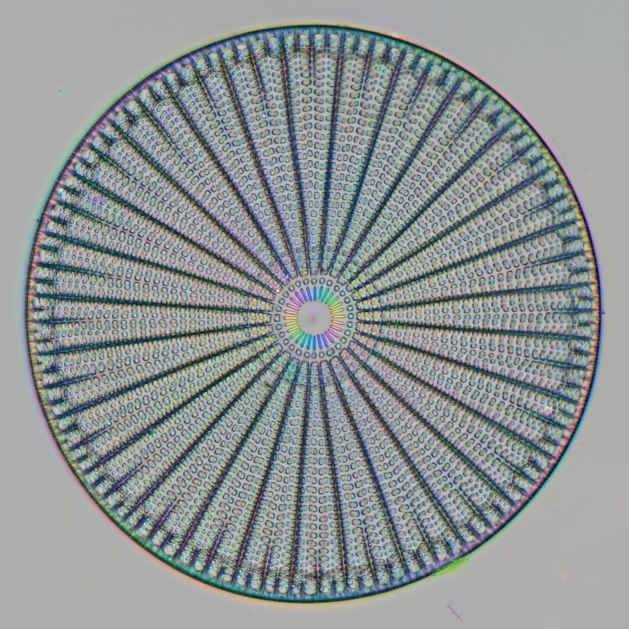
And one more: Shribak received an Honorable Mention in the 2022 EVIDENT Image of the Year Award, sponsored by Olympus Life Sciences (now called Evident). He was recognized for his image of a diatom, a unicellular organism.

Congratulations!